


Patellar Instability
What is Patellar Instability?
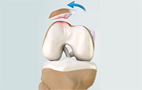
Patellar (kneecap) instability results from one or more dislocations or partial dislocations (subluxations). Patella is the small piece of bone in front of the knee that slides up and down the femoral groove (groove in the femur bone) during bending and stretching movements. The ligaments on the inner and outer sides of patella hold it in the femoral groove and avoid dislocation of patella from the groove. Any damage to these ligaments may cause patella to slip out of the groove either partially (subluxation) or completely (dislocation). This misalignment can damage the underlying soft structures such as muscles and ligaments that hold the kneecap in place. Once damaged, these soft structures are unable to keep the patella (kneecap) in position. Repeated subluxation or dislocation makes the knee unstable and the condition is called as knee instability.
Symptoms of Patellar Instability
Patients with knee instability experience different signs and symptoms such as:
- Pain, especially when standing up from a sitting position
- Feeling of unsteadiness or tendency of the knee to “give way” or “buckle”
- Recurrent subluxation
- Recurrent Dislocation
- Severe pain, swelling and bruising of the knee immediately following subluxation or dislocation
- Visible deformity and loss of function of the knee often occurs after subluxation or dislocation
- Sensation changes such as numbness or even partial paralysis can occur below the dislocation because of pressure on nerves and blood vessels
Causes of Patellar Instability
Various factors and conditions may cause patellar instability. Often a combination of factors can cause this abnormal tracking and include the following:
- Anatomical defect: Flat feet or fallen arches and congenital abnormalities in the shape of the patella bone can cause misalignment of the knee joint.
- Abnormal “Q” Angle: The “Q” angle is a medical term used to describe the angle between the hips and knees. The higher the “Q” angle, such as in patients with Knock Knees, the more the quadriceps pull on the patella causing misalignment.
- Patellofemoral Arthritis: Patellar misalignment causes uneven wear and tear and can eventually lead to arthritic changes to the joint.
- Improper Muscle Balance: Quadriceps, the anterior thigh muscles, function to help hold the kneecap in place during movement. Weak thigh muscles can lead to abnormal tracking of the patella, causing it subluxate or dislocate.
Diagnosis of Patellar Instability
Your surgeon diagnoses the condition by collecting your medical history and physical findings. He may also order certain tests such as X-rays, MRI or CT scans to confirm the diagnosis.
Treatment of Patellar Instability
Treatment for instability depends on the severity of condition and based on the diagnostic reports. Initially your surgeon may recommend conservative treatments such as physical therapy, use of braces and orthotics. Pain relieving medications may be prescribed for symptomatic relief. However, when these conservative treatments yield unsatisfactory response surgical correction may be recommended.
Considering the type and severity of injury surgeon decides on the surgical correction. A lateral retinacular release may be performed where your surgeon releases, or cuts, the tight ligaments on the lateral side (outside) of the patella enabling the patella to slide more easily in the femoral groove.
Your surgeon may also perform a procedure to realign the quadriceps mechanism by tightening the tendons on the inside or medial side of the knee.
If the misalignment is severe tibial tubercle transfer (TTT) will be performed. This procedure involves the surgeon removing a section of bone where the patellar tendon attaches on the tibia. The bony section is then shifted and properly realigned with the patella and reattached to the tibia with two screws.
Following the surgery rehabilitation program may be recommended for better outcomes and quicker recovery.